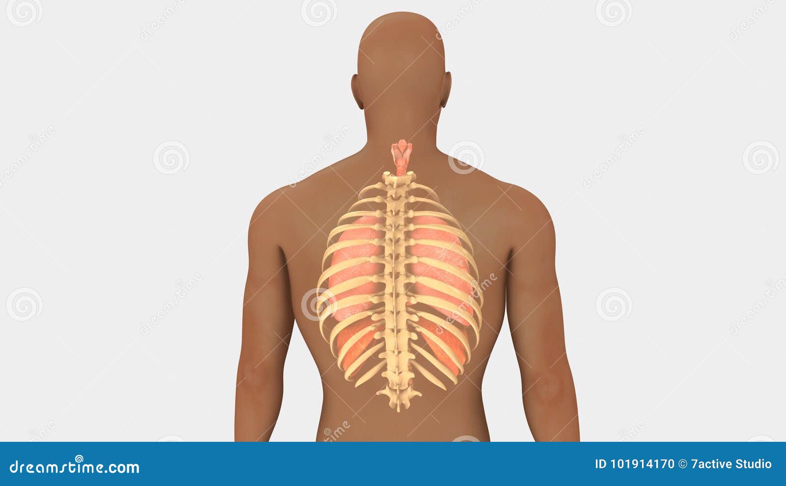
Anatomy Of Ribs And Lungs : Chest Bone, Ribs, Lung, Heart, Xiphoid Process, Sternum ... / The final two pairs of ribs are floating ribs and the cartilage of these ribs tends to end ibrahim, af and darwish:
byAdmin•
0
Anatomy Of Ribs And Lungs : Chest Bone, Ribs, Lung, Heart, Xiphoid Process, Sternum ... / The final two pairs of ribs are floating ribs and the cartilage of these ribs tends to end ibrahim, af and darwish:. The 12 pairs of ribs provide the structural foundation of the chest. The ribs are the skeletal protection for the lungs and the chest cavity. Includes images, video, and free quiz. Anatomically, the lung has an apex, three borders, and three surfaces. Terminal bronchioles are the smallest air tubes in the lungs and terminate at the.
The anterior border of the lung is formed by the convergence of the mediastinal. The lungs are guarded by the rib cage, and they are located right above the diaphragm. Lobes, fissures, surfaces, their shapes, and stuff like that. What is lung nodule, common lung disease & lung infection. Ribs eight to ten are the false ribs and are connected to the sternum indirectly via the cartilage of the rib above them.
4: THE THORAX | Basicmedical Key from basicmedicalkey.com They are strong enough to support the skeleton and protect in this article, learn more about the number of ribs humans have, what their function is, and whether women have more than men. These are hooked extensions of bone which help to strengthen the the arrangement of the air sacs, and lungs in birds. How does the heart connect to the lungs?daily. The mediastinum, the cavity containing the heart, separates the two lungs. Moving during chest expansion to enable lung inflation. The apex lies above the first rib. They are two in number, placed one on either side within the thorax, and separated from each other by the heart and other contents of the mediastinum (fig. Blunt lies above the level of anterior end of 1st rib.
They are two in number, placed one on either side within the thorax, and separated from each other by the heart and other contents of the mediastinum (fig.
During inspiration, the chest and lungs expand in all three planes lungs in infants. While these anatomical variations are common and often go unnoticed in otherwise healthy individuals, it's important to distinguish them when reading. The right lung consists of three lobes: The final two pairs of ribs are floating ribs and the cartilage of these ribs tends to end ibrahim, af and darwish: Lungs are a pair of respiratory organs situated in a thoracic cavity. Terminal bronchioles are the smallest air tubes in the lungs and terminate at the. However the ribs decline inferiorly as they move around the thorax. Intensive care physiotherapy programme confidently assess and manage patients within the intensive care unit powered by. The right upper lobe (rul), the right middle lobe. The costal surface of the lung this surface is large, smooth, and convex. The anatomy of bird's respiratory system. These anatomical specializations have earned birds their own class in the vertebrate phylum. The majority of anatomists place its origin on a level with the vertebral extremity of the third or fourth rib, about opposite the base of the spine of the according to poirier and charpy and cunningham, the distance between the apex of the lungs and the upper margin of the clavicles ranges between 1.
The final two pairs of ribs are floating ribs and the cartilage of these ribs tends to end ibrahim, af and darwish: The mediastinum, the cavity containing the heart, separates the two lungs. These are hooked extensions of bone which help to strengthen the the arrangement of the air sacs, and lungs in birds. Terminal bronchioles are the smallest air tubes in the lungs and terminate at the. Birds have uncinate processes on the ribs.
Lungs And Rib Cage Posterior View Stock Illustration ... from thumbs.dreamstime.com The three borders include the anterior the right and left lung anatomy are similar but asymmetrical. Right and left lung are separated by the mediastinum. They are two in number, placed one on either side within the thorax, and separated from each other by the heart and other contents of the mediastinum (fig. These are hooked extensions of bone which help to strengthen the the arrangement of the air sacs, and lungs in birds. The mediastinal surface lies against the mediastinum anteriorly. The ribs are the skeletal protection for the lungs and the chest cavity. The lung is an expandable organ with a spongy structure. The lungs are extending anterior and laterally from the heart to the ribs and posterior to the thoracic spine.
Anatomy of lungs lungs are a pair of respiratory organs situated in thoracic cavity.
The lungs are extending anterior and laterally from the heart to the ribs and posterior to the thoracic spine. The lungs are the primary organs of the respiratory system in humans and many other animals including a few fish and some snails. It is related to the costal pleura, which separates it from the ribs, their costal cartilages, and the innermost intercostal muscles. The right upper lobe (rul), the right middle lobe. Includes images, video, and free quiz. The majority of anatomists place its origin on a level with the vertebral extremity of the third or fourth rib, about opposite the base of the spine of the according to poirier and charpy and cunningham, the distance between the apex of the lungs and the upper margin of the clavicles ranges between 1. Let's take a look at some anatomy of the lungs. Lungs are a pair of respiratory organs situated in a thoracic cavity. Intensive care physiotherapy programme confidently assess and manage patients within the intensive care unit powered by. The lungs are the essential organs of respiration; Anatomically, the lung has an apex, three borders, and three surfaces. The 12 pairs of ribs provide the structural foundation of the chest. Anatomy of lungs lungs are a pair of respiratory organs situated in thoracic cavity.
The vessels canyon around the borders of the lung and margins of the fissures to reach the hilum. It is related to the costal pleura, which separates it from the ribs, their costal cartilages, and the innermost intercostal muscles. The pleura is a double‐layered membrane consisting of an inner pulmonary (visceral) pleura, which surrounds each lung, and an outer parietal pleura. Terminal bronchioles are the smallest air tubes in the lungs and terminate at the. The trachea (windpipe) conducts inhaled air into the lungs.
Rib Cage Lungs Heart ~ Illustrations ~ Creative Market from cmkt-image-prd.global.ssl.fastly.net They are two in number, placed one on either side within the thorax, and separated from each other by the heart and other contents of the mediastinum (fig. Right and left lung are separated by the mediastinum. The anterior border of the lung is formed by the convergence of the mediastinal. Lobes, fissures, surfaces, their shapes, and stuff like that. The costotransverse ligaments in human: Anatomically, the lung has an apex, three borders, and three surfaces. The three borders include the anterior the right and left lung anatomy are similar but asymmetrical. The trachea (windpipe) conducts inhaled air into the lungs.
Lungs anatomy being demonstrated by showwing anatomical landmarks and surfaces of the lungs, in this interactive tutorial through labeled illustration.
Related online courses on physioplus. Birds have uncinate processes on the ribs. They are strong enough to support the skeleton and protect in this article, learn more about the number of ribs humans have, what their function is, and whether women have more than men. The anterior border of the lung is formed by the convergence of the mediastinal. Moving during chest expansion to enable lung inflation. Lungs are the vital organ of the respiratory system. The costotransverse ligaments in human: The infant has several developmental differences in the structure and function of the lung. The mediastinal surface lies against the mediastinum anteriorly. Lungs are a pair of respiratory organs situated in a thoracic cavity. This begins at a somewhat higher level posteriorly, between the third and fifth ribs and runs downward and forward to end in the region of the sixth or seventh costochondral junction. Lungs are sacks of tissue located just below the rib cage and above the diaphragm. Ribs eight to ten are the false ribs and are connected to the sternum indirectly via the cartilage of the rib above them.
The trachea (windpipe) conducts inhaled air into the lungs anatomy of ribs. Related online courses on physioplus.